
The most common locations are the extremities. Unlike classic spindle cell/pleomorphic lipomas, ASPLT has a wide anatomical distribution and the clinical differential diagnosis depends on the involved location. This entity can affect both sexes, with a slight male predominance, including mostly middle-aged adults with a peak in the sixth decade, likewise our patient. Our case represents an atypical pleomorphic lipomatous tumor characterized by the presence of floret-like multinucleated giant cells. As result, there is a wide range of microscopic appearance of ASPLT due to the extremely variable proportions of spindle cells, adipocytes, lipoblasts, and extracellular matrix. At the other extreme, they may be significantly more cellular, with less extracellular matrix.

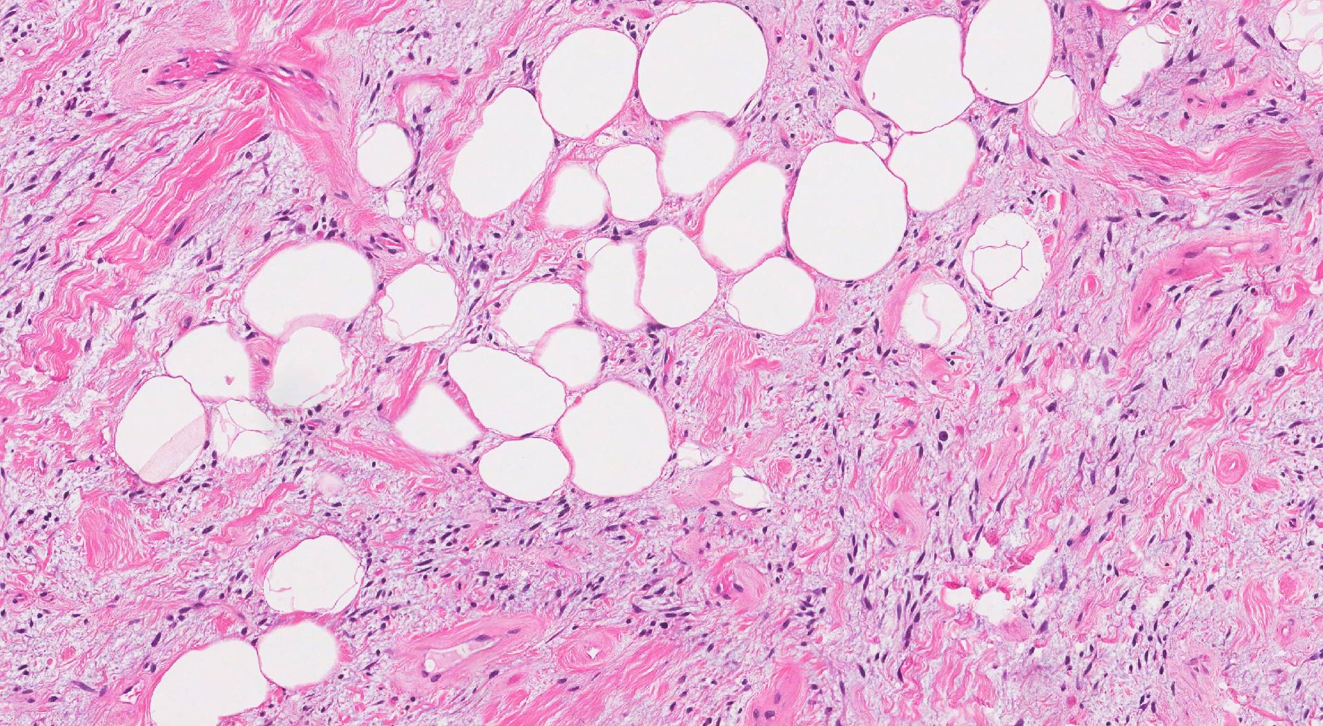
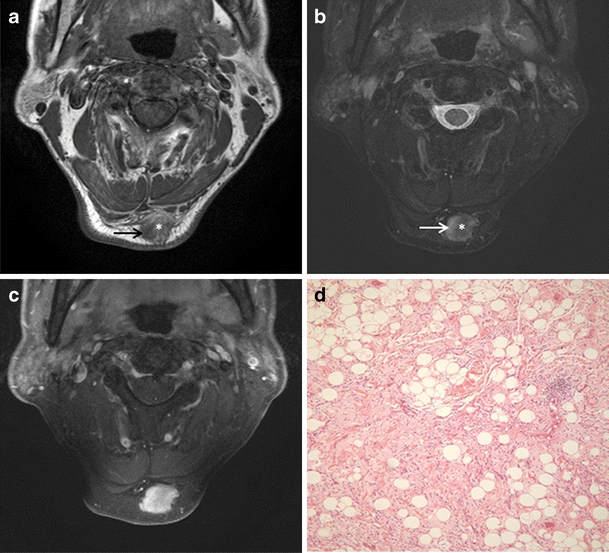
The cellularity of the tumors is the basis of the spectrum. At one end, tumors can be paucicellular with few atypical spindle cells and abundant extracellular matrix. ĪSPLT is a morphologic spectrum of benign adipocytic neoplasms. Since then, other works have given strength to this theory, representing now a newly established diagnostic entity. proposed the term “atypical spindle cell lipoma” for the first time in 2010, suggesting that these neoplasms most likely represent an independent entity closely related to spindle cell lipoma rather than a morphologic variant of atypical lipomatous tumor/well-differentiated liposarcoma. Recently, WHO published the 5 th edition of soft-tissue tumors classification which included a new group, the atypical spindle cell/pleomorphic lipomatous tumor (ASPLT). Although they are mostly lipomas or angiolipomas, other rare entities can arise and raise doubts. Based on these findings, an atypical pleomorphic lipomatous tumor was diagnosed.Ĭutaneous tumors with adipocyte differentiation are frequently excised by surgeons. On immunohistochemistry, the tumor was stained for CD34, S100, and MDM2 (focal-weak), whereas CDK4 expression was absent (Figure 4). Sclerosing areas were not disclosed (Figure 3). Microscopically, a variable amount of atypical bland spindle cells and mature adipocytes were seen, with multinucleated floret-like cells in a myxoid stroma with ropey collagen bundle cells. Grossly, it was a subcutaneous nodular non-capsulated solid lesion, multilobulated, well-circumscribed, greyish-yellowish, without necrotic areas (Figure 2). There were no complications related to the procedure. Based on clinical and image findings, it was decided to perform an excisional biopsy.ĭespite the apparent benign characteristics, the lesion was surgically removed along with the surrounding adipose tissue, preserving the margins. An MRI described a “focal subcutaneous lesion with nodular morphology of 4.7 cm and no malignancy features”. The patient was referred to the General Surgery department by a urologist, with suspicion of a soft-tissue tumor.


 0 kommentar(er)
